Platelet-rich fibrin: a versatile regenerative treatment for dogs, cats and horses

Regenerative medicine can do far more than we currently use it for in small animal clinics. Platelet-rich fibrin (PRF) is a 100% autologous product that can be prepared quickly in the clinic and used for dental treatment, wound care, joint treatment and corneal ulcers, among other things. The optimal mix of platelets, leucocytes, fibrin and growth factors supports and secures rapid healing.

Read about treatment areas and PRF preparation on this page, sign up for our PRF course for veterinarians, or buy a PRF setup for your clinic.

PRF helps wounds heal faster

PRF is used in human medicine and increasingly in veterinary medicine, where several studies have shown improved wound healing compared with conventional treatment. A 2024 study (Soares et al.) showed that naturally occurring wounds in dogs responded well to treatment with PRF membranes, and that all included wounds achieved complete healing.

A 2023 study (Changrani-Rastogi et al.) showed that naturally occurring wounds in cats healed faster with PRF than in the control group. In the group of cats treated with PRF, the wounds had contracted 94% in 14 days, whereas only 76% contraction was seen in the control group.

PRF can be particularly interesting in chronically infected or complicated wounds where healing is delayed, as well as in larger skin defects or wounds with exposed underlying tissue. The aim is to support the formation of granulation tissue and create optimal conditions for the further healing process.

Join a PRF course

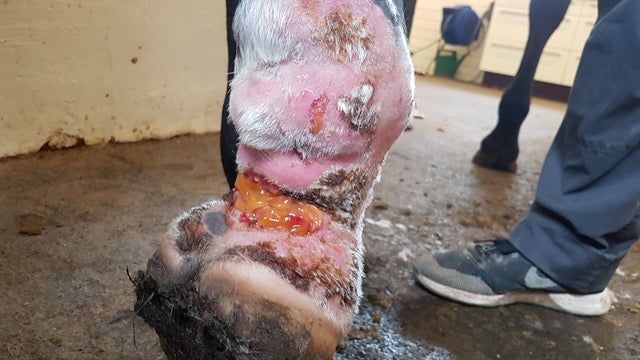
Wound healing with PRF

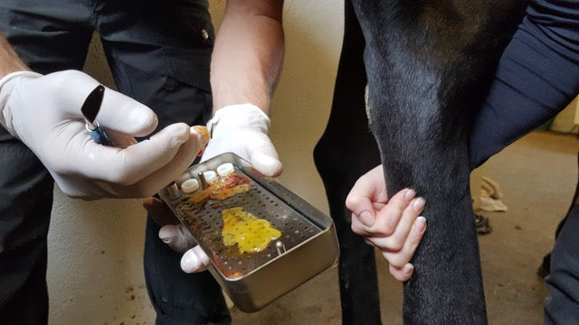
Preparation of PRF

PRF membranes are made quickly on site, drawing enough blood to cover the intended area.

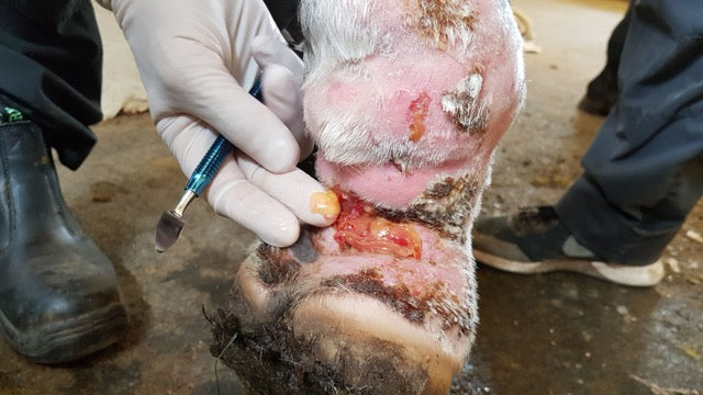
Add a membrane to the wound

The PRF membrane is applied to the wound after debridement. Shown here is a laceration on a horse’s pastern, which often causes complications.

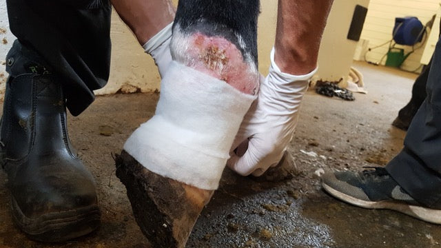
Standard dressing

The wound is dressed as usual after PRF is applied. PRF treatment is typically repeated once or twice a week as needed while healing is monitored closely.

PRF supports healing after dental treatments

PRF is a promising tool in veterinary dentistry, where it can be used to support the healing of both soft tissue and bone after dental treatments. Studies have shown that PRF can stimulate the regeneration of periodontal tissue in dogs, including the formation of new bone, cementum and ligament tissue in periodontal defects. The results show that PRF, in addition to promoting soft tissue healing, can support the healing of tissue that otherwise has limited regenerative capacity.

PRF is also relevant in connection with tooth extractions. In a 2022 study (Liu et al.), the use of PRF resulted in improved early gingival healing after extraction compared with control groups.

PRF is therefore an obvious option for larger extraction sockets, surgical extractions, periodontitis, oronasal fistulas or patients where rapid soft tissue healing is desired. PRF is placed in extraction sockets, periodontal defects and other surgical sites, where it acts as a biological scaffold and contributes growth factors that support early tissue healing.

Join a PRF course

Dental treatment with PRF

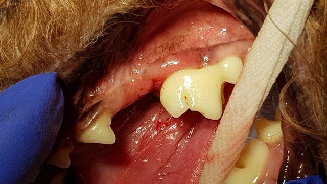
The defect is assessed

Here is a deep pocket at risk of attachment loss. Tooth preservation is judged possible with PRF.

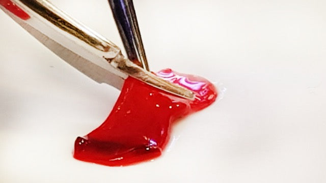
Preparation of PRF

The patient’s blood is drawn and a PRF membrane is formed and trimmed to fit the intended area.

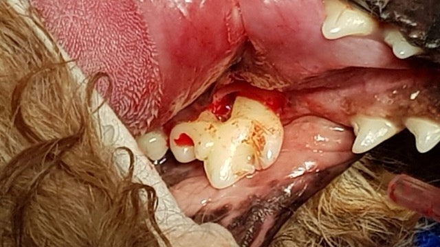
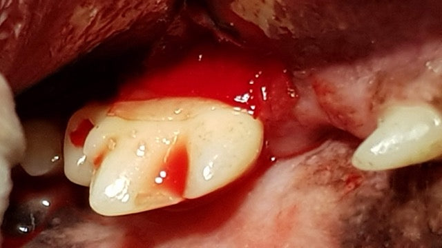
Add a membrane to the defect

The PRF membrane is applied to the gingiva and around the root. Healing is monitored closely and radiographs are taken later.

Liquid PRF improves healing of joint cartilage

PRF was originally developed as a clot that could be shaped into membranes and used to support wound healing and tissue regeneration. Later, a liquid, injectable version was developed.

The liquid form makes PRF particularly suitable for treating joints and tendons, as it can be injected directly into the desired area. After injection, PRF gradually coagulates and forms a fibrin network that releases growth factors and cytokines over several days. This supports the body's natural healing processes and dampens local inflammation.

A 2014 study (Kazemi et al.) showed that articular cartilage treated with PRF achieved significantly better healing than untreated cartilage. A larger 2023 clinical study (Cheeva-Akrapan et al.) in patients with osteoarthritis likewise showed that liquid PRF produced improvements in pain and function and supported the use of the treatment. In addition, a 2025 study (Najeb et al.) in donkeys with superficial digital flexor tendonitis showed that liquid PRF promoted healing of the tendon injury.

In practice, liquid PRF can be prepared directly from the patient's own blood during the consultation and injected intra-articularly or peritendinously. The treatment can therefore be a supplement to existing treatment of dogs with osteoarthritis, arthritis or tendon injuries, where the aim is to support tissue healing and reduce inflammation using the patient's own biological growth factors.

Join a PRF course

Liquid PRF for joints

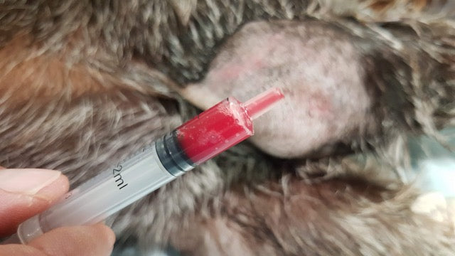
Preparation of liquid PRF

Liquid PRF is prepared from the patient’s own blood and drawn into a syringe. It must be used promptly, as coagulation begins after about 15 minutes.

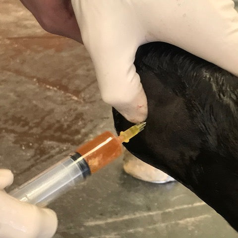
Joint injection in the tarsal joint

Liquid PRF used here for injection into the tarsal joint of a horse.

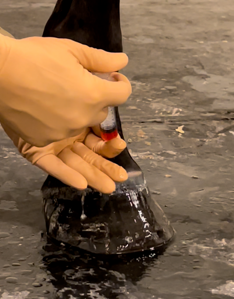
Joint injection in the pastern joint

Liquid PRF used here for injection into the pastern joint of a horse.

PRF promotes tissue healing in the eye

The healing of corneal ulcers can be challenging, as the cornea is avascular and therefore depends on the surrounding tissue for the supply of oxygen, nutrients and growth factors. In larger or deeper ulcers, this limited blood supply can prolong the healing process. PRF can create a more optimal environment for tissue healing by locally supplying a fibrin matrix with platelets, leukocytes and growth factors.

The PRF membrane acts both as a biological scaffold that supports the migration of epithelial cells, and as a depot that gradually releases growth factors over several days. This can stimulate repair and contribute to a more controlled inflammatory response, which overall can promote healing and reduce the risk of further tissue damage.

Studies in both dogs and horses have shown promising results in the treatment of corneal ulcers with PRF. Two studies published in 2023 (Baadsgaard Bruun et al.) (Hosny et al.) conclude that PRF is a safe and effective supplementary treatment method that can shorten healing time, improve wound healing and reduce the risk of complications compared with conventional treatment alone.

PRF can be produced as a membrane that is adapted to the size of the corneal ulcer and placed directly on the lesion. The membrane is typically fixed with sutures or protected with a temporary tarsorrhaphy so that it is held stable over the ulcer during healing. In this way PRF acts both as a biological dressing and as a local source of growth factors that support the tissue's natural regeneration process.

Join a PRF course

Healing corneal ulcers with PRF

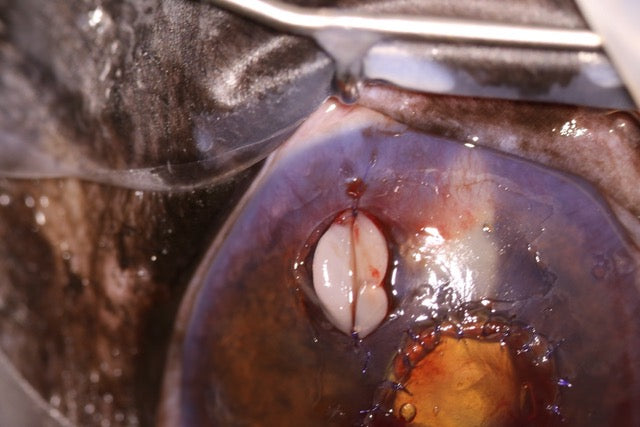
PRF membranes are trimmed to fit

PRF membranes are made from the patient’s own blood and trimmed to the size of the wound.

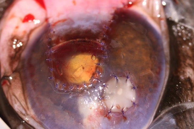
Membrane sutured in place

PRF membranes are sutured onto the cornea.

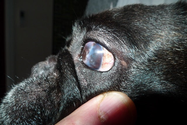
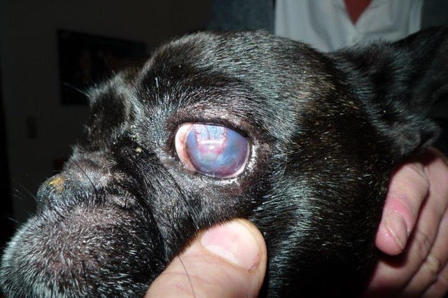
Follow-up

Healing is monitored closely. Here is the dog’s eye 10 days after PRF treatment began.

PRF course: PRF in veterinary medicine

The route to faster healing, fewer complications and greater patient confidence

Learn more about PRF on our course on 26 November 2026

Read more

How is PRF made?

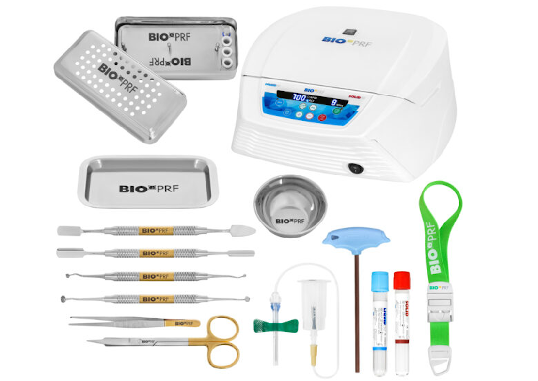
PRF is easily prepared in the clinic with a few instruments and the patient’s own blood. Different protocols are followed depending on whether a membrane or liquid PRF is required.

Preparing a PRF membrane in 3 steps:

1. Centrifuging the blood

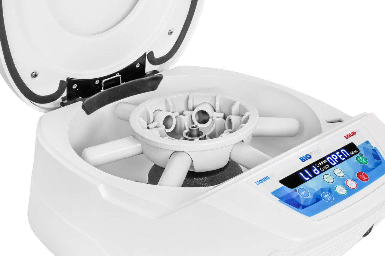
After blood collection into glass tubes without anticoagulant, the sample is centrifuged. The optimal angle is achieved with a horizontal centrifuge.

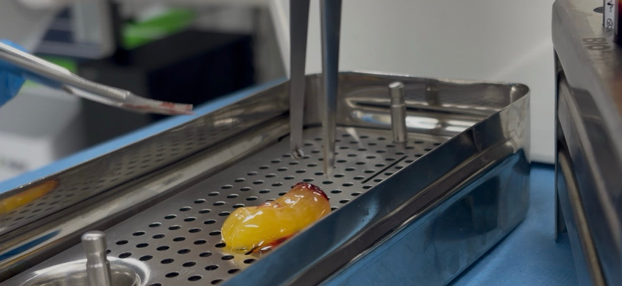
2. Removing the PRF clot

During centrifugation a fibrin clot (PRF clot) forms between the cellular blood fraction and the plasma. The clot is then carefully removed from the tube and can be separated from the red blood cells with scissors or tweezers.

3. Application of PRF

The PRF can now be used directly as a biological implant or compressed into a membrane, depending on the intended use.

Preparing liquid PRF in 3 steps:

1. Centrifuging the blood

Liquid PRF is prepared in plastic tubes without anticoagulant, and the sample is spun in a horizontal centrifuge.

2. Collecting liquid PRF

The top millilitres of the sample are discarded and the remaining plasma is drawn into a syringe (without taking red blood cells).

3. Application of liquid PRF

The PRF can now be injected into joints or other areas as needed. After 15 minutes a fibrin scaffold starts to form.